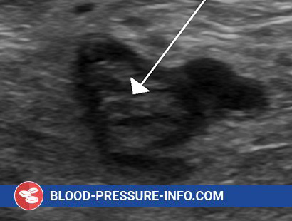
What is Thrombophilia Associated with Antithrombin III Deficiency?
Genetically determined (primary) forms This group includes all types of hereditary insufficiency of the main physiological anticoagulant, antithrombin III, with a pronounced tendency to multiple recurrent thromboembolisms and organ infarctions.
Causes of Thrombophilia Associated with Antithrombin III Deficiency
Thrombophilia is an autosomal dominantly inherited disease with different penetrance of the pathological gene. It was first described as an independent disease in 1965.
In most cases, thrombophilia is characterized by a violation of the synthesis of AT III, in which the functional activity of this anticoagulant and the content of its antigen in the plasma are equally reduced.
Much less common are antigen-positive forms, in which the low functional activity of AT III is combined with a normal or almost normal content of its antigen in the plasma, which indicates the preserved production in the body of “abnormal”, not functioning as an anticoagulant of the AT III molecule.
The prevalence of genetically determined thrombophilia has not been precisely established, since mainly only the most severe (clinically pronounced) forms of it are detected. According to tentative sample data, their frequency ranges from 1: 5000 to 1: 2000 families, but among patients with venous thrombosis and pulmonary embolism, AT III deficiency is detected in 2-3% of cases. Among all hereditary coagulopathies, including hemophilia, AT III deficiency is the most common pathology. Some observations show that, along with clinically clearly expressed severe thrombophilia, erased and inapparent forms of the disease are widespread. With them, spontaneous thromboembolism is rare or absent, but it easily occurs under the influence of additional provoking factors – surgical interventions, trauma, pregnancy and childbirth, immobilization of the limbs.
Pathogenesis during Thrombophilia Associated with Antithrombin III Deficiency
The clinical picture of thrombophilia consists of recurrent venous and arterial thrombosis of different localization, embolism in the basin of the pulmonary artery and other vessels, organ infarctions (lungs, myocardium, brain, kidneys), developing at a relatively young age and characterized by more or less pronounced resistance to heparin therapy.
The more significant the deficiency of AT III, the earlier the disease manifests itself and the more severe it is.
The most severe are the homozygous forms of the disease, caused by its double inheritance from both parents, which are most common in those regions where consanguineous marriages are practiced. In such a situation, the level of AT III in the child’s plasma does not exceed 2-3%, which leads to the early development of fatal thromboembolism.
In the clinically expressed heterozygous form of the disease, the level of AT III in plasma ranges from 20 to 35%, the thrombosembolic syndrome usually debuts between the ages of 16 and 35 years.
The first thrombosis with a deficiency of AT III usually occurs spontaneously or under the influence of provoking factors – significant physical exertion, sudden cooling or overheating, trauma and surgical interventions, during pregnancy, especially complicated by toxicosis, after childbirth.
The onset of the disease can also be associated with medical interventions that increase the thrombogenic potential of the blood, the intake of synthetic progestins (contraceptives, infectiousdin, mestranol), intravenous administration of aminocaproic acid and plasma coagulation factor concentrates (especially PPSB), vascular catheterization, and arteriovenous shunts.
The first most often thrombosed are the saphenous and deep veins of the lower extremities and pelvic region, but the disease can begin with thrombosis of any localization. Thrombotic episodes are repeated with greater or lesser frequency (light periods last from several days to weeks and months), and more and more new vessels are involved in the process. Often, vessels of different localization and caliber are thrombosed at the same time or almost simultaneously, which confirms the systemic nature of the disease.
In general, thrombosis of different localization in thrombophilia creates a variegated palette of organ manifestations that have a common foundation – primary AT III deficiency.
Along with severe forms of the disease (homo- and heterozygous), it is advisable to isolate a borderline form with an AT III level in the range of 45 to 65% and a potential one with an AT III level between 65 and 85% in the interthrombotic (“cold”) period, which does not require treatment.
In the borderline form, spontaneous thrombosis is rare and few in number, appearing at a later age than in severe thrombophilia. Sometimes they are completely absent, but naturally arise under the influence of additional thrombogenic effects and diseases. After eliminating these influences, the thrombotic process does not continue, as in a severe form, but fades away or gives very rare relapses. In such patients, it is very easy to provoke thrombosis – the veins are often thrombosed after conventional intravenous injections, and each injection of a needle into the vein poses a risk of thrombosis. As a result of the treatment of various antecedent diseases, all veins are clogged and further intravenous administration of drugs becomes impossible. The preservation of veins in patients suffering from both AT III deficiency and other types of thrombophilia is becoming more and more important, which should be taken into account when planning therapeutic interventions. In particular, it is important to avoid multiple intravenous injections, carefully consider the thrombotic history, including blockage of veins after injections, difficulty in obtaining blood from a vein due to rapid clotting in the needle.
In a potential form of the disease, thrombosis occurs only when the AT III level near the lower limit of the norm (75–85%) decreases under the influence of any additional influences. Such an additional decrease in the level of AT III naturally occurs on the 3-5th day after major operations, with toxicosis of pregnancy and after childbirth, with septicemia and shock conditions, bleeding, injections of large doses of unbalanced, i.e., not containing AT III, hemopreparations ( PPSB, cryoprecipitate, fibrinogen), due to the introduction of synthetic progestins and a number of other reasons, including intensive or prolonged use of heparin, under the influence of which the catabolism of AT III is enhanced.
Thus, potential thrombophilia should be considered as a pre-thrombotic condition in which a real threat of thromboembolism arises under the influence of factors that further reduce the level of AT III in plasma.
It should be emphasized that borderline and potential forms of thrombophilia cannot be considered “mild” or “latent”, since any episode of thromboembolism in these forms can be fatal or cause disability (due to paralysis, organ infarctions).
In connection with the seriousness of the prognosis of patients with all types of thrombophilia, it is necessary to actively identify and provide them with dispensary observation. It is important to conduct a laboratory examination of all relatives of identified patients.
Diagnosis of Thrombophilia Associated with Antithrombin III Deficiency
Diagnosis is based on a thorough study of the individual and family thrombotic history, taking into account not only obvious thromboembolism, but also such minor manifestations as vein thrombosis after injections, the appearance of asymmetric edema of the extremities in the evening or after several hours of sitting, the feeling of “sitting out” is always the same leg, thrombosis during pregnancy, after surgery and trauma, as well as due to the use of hormonal contraceptives.
The diagnosis becomes more likely when a sharp weakening of the anticoagulant effect of heparin is detected, although resistance to heparin may be due to other reasons.
The diagnosis is confirmed only by quantitative determination in plasma of AT III, for which functional coagulation, amidolytic and immunological methods are used.
Thrombophilia due to molecular abnormalities of AT III are not diagnosed by immunological methods, since the level of the AT III antigen remains normal. The results of functional determinations can be distorted by heparin therapy, the presence in the plasma of a large amount of fibrinogen cleavage products.
Also, a decrease in AT III in plasma can develop secondarily, as a result of massive thrombosis or in severe liver damage, disseminated intravascular coagulation, nephrotic syndrome, systemic vasculitis.
The diagnosis of hereditary thrombophilia is considered proven only when a low level of AT III remains stable in the interthrombotic period, in the absence of background diseases and outside of anticoagulant and transfusion therapy.
The second important laboratory characteristic of thrombophilia is a sharp weakening of the anticoagulant effect of heparin both in in vitro experiments and with intravenous administration. This phenomenon is most clearly detected using a serial heparin-thrombin test.
Particular attention is paid to patients with a history of thrombotic episodes, including those after intravenous interventions, as well as blood relatives of identified patients with thrombophilia. Patients are subject to the same examination before installing a vascular catheter, arteriovenous shunting, prosthetics of vessels and heart valves and many other interventions on the vessels and heart.

